
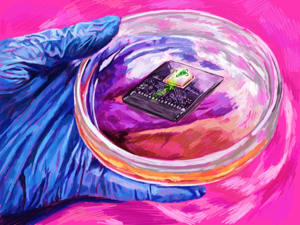
The processes behind skin cells closing wounds and tumor cells spreading throughout the body both have to do with cell movement, but tracking that movement can be difficult and often requires using fluorescent dyes, which can have negative side effects. Brown researchers have developed a new imaging technology that uses electrical measurements to track individual cells’ movement.
“For the past several years in the Rosenstein Lab, we have been interested in the goal of building a chip to track cells by mapping their electrical and chemical properties, instead of using light,” Jacob Rosenstein, an author of the study and associate professor of engineering, wrote in an email to The Herald.
The sensors measure capacitance — the amount of charge that can be stored between two conductors — to observe cell movement, Rosenstein wrote.
According to Rosenstein, by avoiding the use of fluorescence dyes and labels, this sensor makes it possible to image a wider range of cells and avoid negative possible health effects from dyes.
The new sensor also covers a much larger area than a traditional optical microscope, which uses light to magnify images. This larger area allows it to collect much more information about the movement and behavior of hundreds of thousands of cells over time, Associate Professor of Engineering and of Pathology and Laboratory Medicine Ian Wong — the principal investigator of the study — wrote in an email to The Herald.
The sensor can observe both individual and collective movement of cells. Some human cell behaviors, “such as swarming or swirling … cannot be understood by only considering individual cells,” Wong wrote.
According to Rosenstein, since the sensors are small, light and relatively inexpensive, they may be useful for wearable medical devices, portable care tools or screening platforms.
“An exciting possibility is to isolate cells from human patients and immediately image them with these sensors to measure how they move,” Wong explained. “This could provide clinically useful information on how invading cancer cells respond to drugs, or how immune cells are activated in response to infection.”
Eric Darling — an associate professor of medical science, engineering and orthopaedics who was not involved in the study — called the technique developed by the paper “highly innovative.” Compared to using fluorescent markers, the technology’s capacitance-based imaging approach provides a “cleaner output to study how cells (and) tissues attach to a surface and move across a two-dimensional field over time.”
The next steps in bettering this technology, Rosenstein wrote, include refining the sensor chips. He added that “there are still opportunities to further improve the resolution, to combine capacitance imaging of cell cultures with more chemically-specific sensing and to apply tools from machine learning and image processing to take full advantage of this new type of sensor data.”
Amrita Rajpal is a senior staff writer covering science and research.


